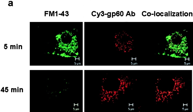
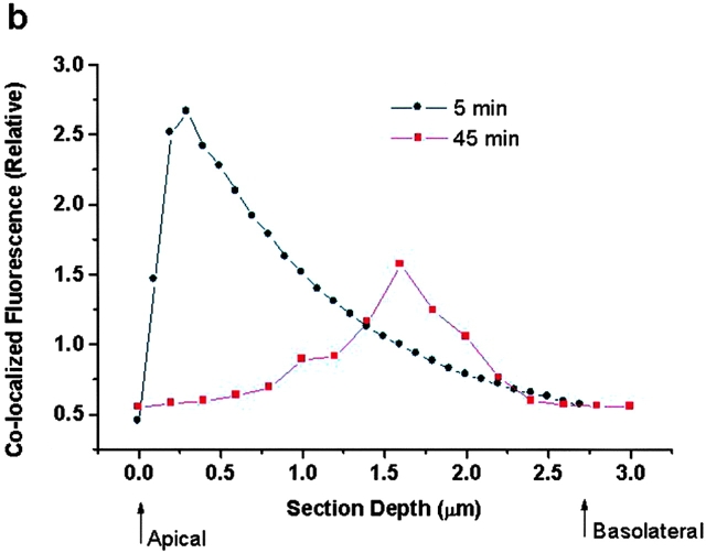
Figure 3.
Colocalization and migration of gp60 and plasmalemma-derived vesicles. (a) Cy3-labeled anti-gp60 Ab was used to fluorescently tag gp60 in BLMVEC incubated in 10 mg/ml BSA. Endosomes were labeled at the same time with 5 μg/ml FM 1-43 to show colocalization of vesicles with gp60. After coincubation with both probes for the indicated times, cell-surface fluorescence was removed by extensive rinsing with pH 5.0 buffer at 4°C. Top row shows confocal images (63× objective), near the luminal cell surface, of early (5 min) intracellular fluorescence because of vesicle marker FM 1-43 (green, left), cy3-labeled anti-gp60 Ab (red, middle), and colocalization image (yellow, right). Bottom row shows albumin cell surface after exocytosis. Note the relative absence of colocalization at 45 min. (b) Plot of migration of gp60-containing vesicles in an endothelial cell. Plot gives relative colocalized fluorescence intensity versus depth of the optical section through the endothelial cell. Peak fluorescent intensity occurred near the luminal cell surface at 5 min after colabeling, whereas the peak is shifted towards the basolateral surface at 45 min. The peak colocalized fluorescence intensity decreased at 45 min compared with 5 min because of exocytosis of FM 1-43 and dilution of its fluorescence in extracellular fluid. Results are representative of five experiments.


