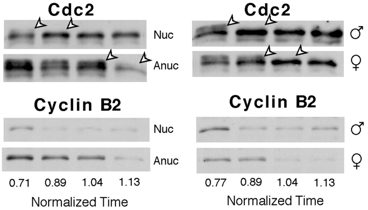
Figure 5.
MPF activation depends on nuclei and centrosomes. Cdc2 and cyclin B2 proteins followed by Western blotting (see Fig. 2) in nucleate (Nuc) and anucleate (Anuc) fragments compared with fragments containing a single male or female pronucleus. The centrosome associated with the male pronucleus organizes a microtubule aster. Cdc2 dephosphorylation was first detectable in fragments containing both nuclei and asters, then in fragments containing the female pronucleus, and finally in anucleate fragments. Low cyclin B2 levels in the last lane of anucleate samples (left blot) partially reflects reduced loading levels (compare with Cdc2 band intensities).

