
Figure 1.
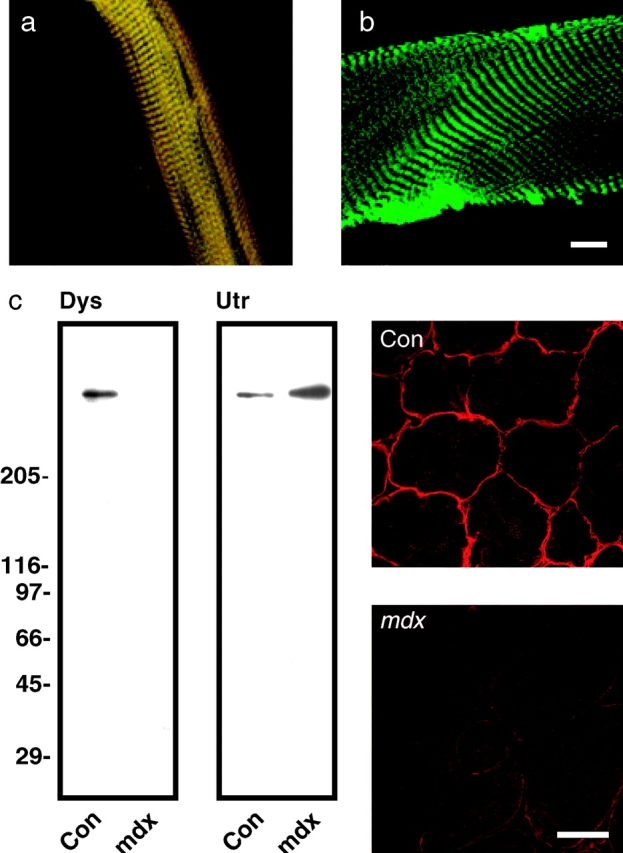
Dystrophin and F-actin colocalize on mechanically isolated sarcolemma in a costameric pattern. Shown is a mechanically isolated sarcolemma (a), or a skinned myofiber (b) both stained with Alexa488-phalloidin (green) and rabbit 2 antiserum to dystrophin (red). Red and green channels were collected simultaneously and areas of coincidence appear yellow. Shown on the left in (c) are immunoblots containing WGA-Sepharose eluates from detergent solubilized control and mdx skeletal muscle membranes stained with rabbit 2 antiserum to dystrophin (Dys), or rabbit 56 antiserum to utrophin (Utr). Shown on the right in c are transverse cryosections of control and mdx skeletal muscle stained with rabbit 2 antiserum to dystrophin. Bars: (b) 10 μm; (c) 50 μm.