
Figure 4.
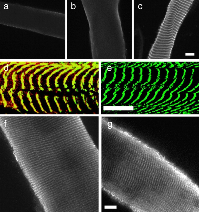
Costameric γ-actin is appropriately expressed and localized in mdx muscle in situ. Shown in a–c are images of sarcolemma from normal mouse muscle stained with antibodies specific to α-sarcomeric (a), β-nonmuscle (b), or γ-actin (c). Shown in d and e are images of control (d), or mdx sarcolemma (e) double stained with monoclonal antibodies to α-actinin (green) and polyclonal antibodies to γ-actin (red). Areas of coincidence between the two probes appear yellow. Shown in f and g are images of fixed and permeabilized myofibers from control (f) and mdx (g) muscle stained with antibodies specific for γ-actin. Bars, 10 μm.