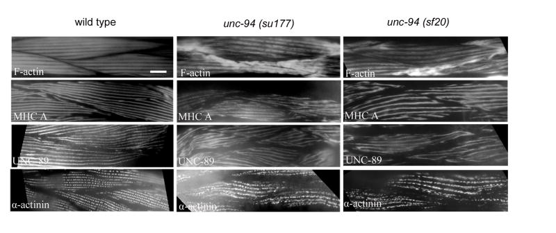
Figure 2.
Immunofluorescent localization of several known sarcomeric proteins in wild type and unc-94 mutant muscle. Antibodies were used to detect MHC A (thick filaments), F-actin (thin filaments), UNC-89 (M-lines), and α-actinin (dense bodies). Each of these proteins shows some degree of mislocalization, in a similar way for both mutant alleles. The most dramatic effect is on the localization of F-actin, with abnormal accumulation near muscle cell / cell boundaries; in most other areas of the cell, I-band organization appears normal. Scale bar represents 10 μm.

