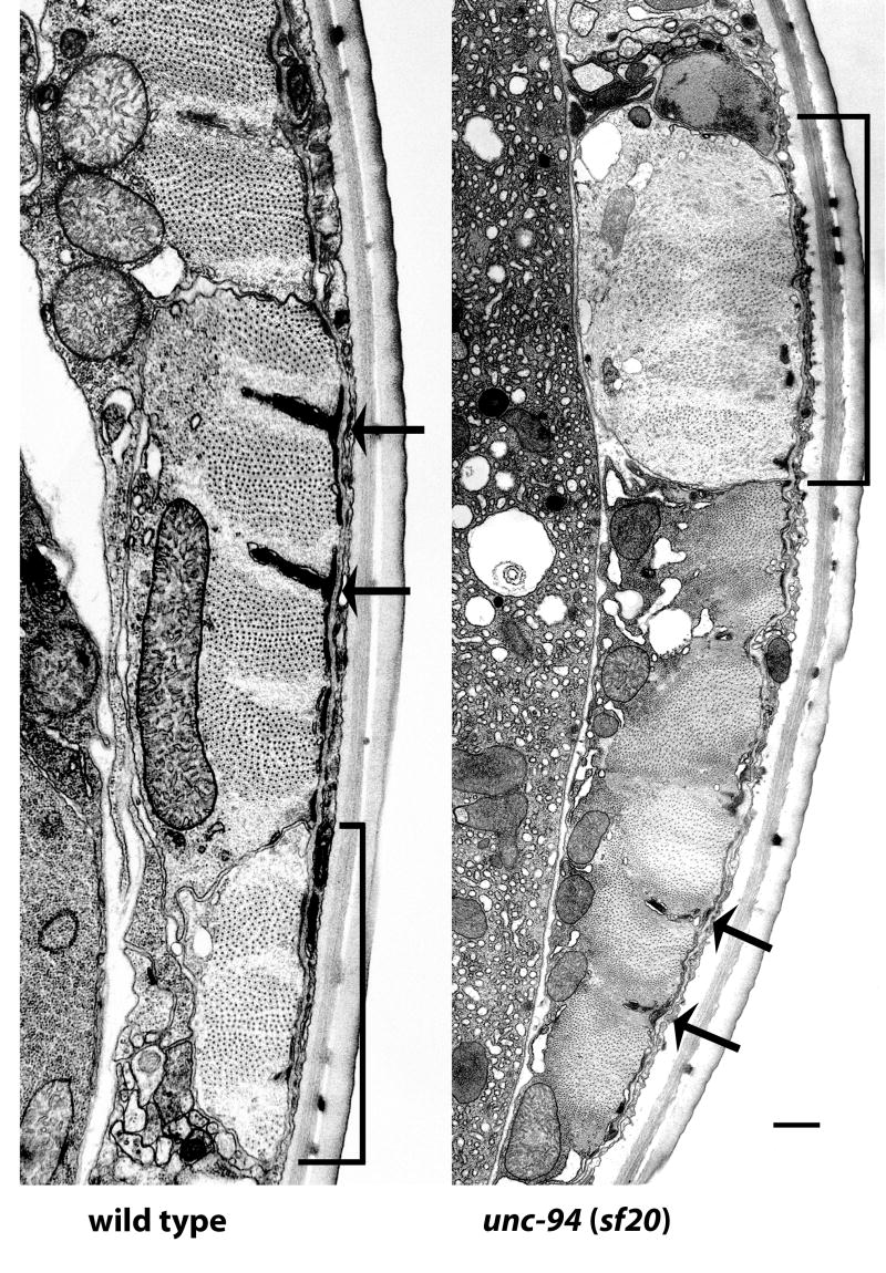
Figure 3.
Electron micrographs of body wall muscle from wild type and from unc-94(sf20). These low power electron micrographs show most of one muscle cell next to a muscle cell process (indicated with a bracket), cut in transverse section. Thick filaments appear as large dots; thin filaments appear as barely discernible thin dots, most prominent around dense bodies. Dense bodies are indicated with arrows. In wild type highly ordered sarcomeres are present, with thin filaments in normal positions, either in I-bands, surrounding dense bodies, or in A-bands associated with thick filaments. Note that in sf20, the muscle cell process is dilated and contains a higher ratio of thin to thick filaments than wild type; also, a small amount of dense body material is found close to the cell membrane. In the adjacent muscle cell, sarcomere organization is better, but dense bodies are irregular. Scale bar, 500 nm.

