
Figure 7.
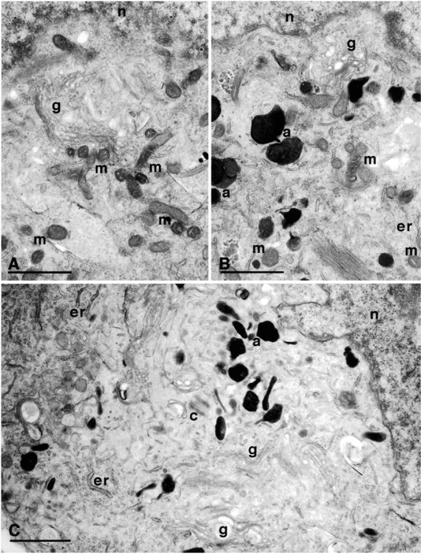

NGF-deprived, BAF-treated 0-DIV neurons lose their mitochondria within 3 d of NGF addition. 0-DIV SCG neurons were fixed for EM after being: (A) treated with NGF for 1 d; (B) NGF-deprived and BAF-treated for 1 d; and (C) NGF-deprived and BAF-treated for 1 d, followed by stimulation with NGF for 3 d. m, mitochondria; g, Golgi; n, nucleus; a, autophagic bodies; c, centriole. Note the complete absence of mitochondria in C. (D) Up to 202 individual cell sections of neurons from each of the three treatments described above were visualized under the EM viewer, and the number of mitochondria per section was counted. Data were sorted into 12 groups (sections containing 0–10 or >10 mitochondria) and plotted as a percentage of neurons containing 0–10 or >10 mitochondria per cell section. Cells maintained with NGF contained 59.2 ± 10.3 mitochondria per section (mean ± SD) and <2 autophagic particles per section, whereas 93/99 sections shown in B contained at least 6 autophagic particles.