
Figure 3.
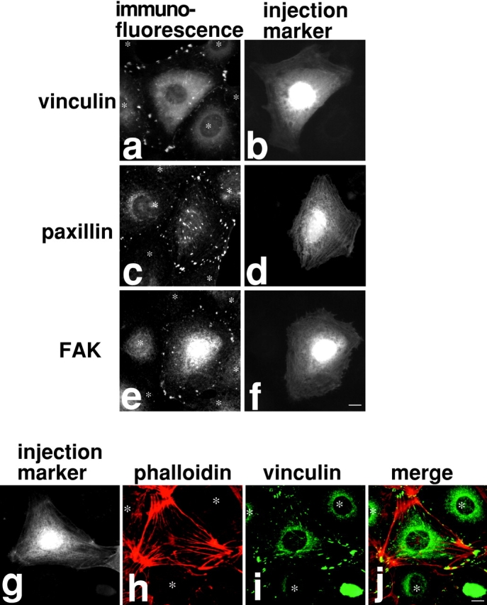
Microinjection of M130Ab induces focal adhesion formation in serum-starved 3T3 cells. a–f: M130Ab was microinjected into serum-starved 3T3 cells as in Fig. 2. Cells were fixed and stained with anti-vinculin antibody (a and b), anti-paxillin antibody (c and d), or anti-FAK antibody (e and f). FITC-dextran was coinjected to identify injected cells (b, d, and f). (g–j) M130Ab-injected cells were double stained with rhodamine-phalloidin (h, red) and anti-vinculin antibody (i, green). Injected cells were detected by anti-rabbit IgG secondary antibody (g). A merged image of rhodamine-phalloidin staining (red) and vinculin localization (green) is shown in j. Asterisks indicated uninjected cells. Bars, 10 μm.