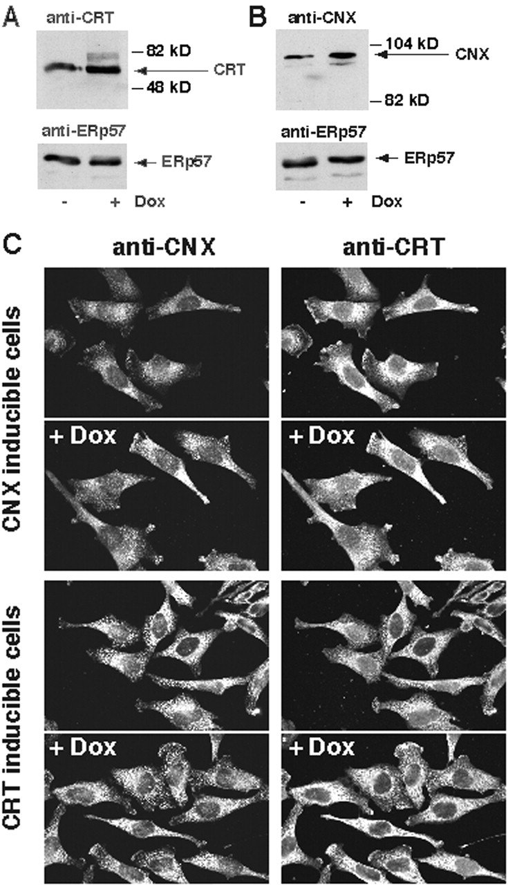
Figure 3.

Expression of calreticulin and calnexin in Tet-On–inducible HeLa cell lines. Overexpression of calreticulin (A, KN1 cells) or calnexin (B, KNX2 cells) was induced by incubation of the cells in the presence of 2 μM Dox for 24 h. Cells were harvested, lysed with RIPA buffer. 10 μg of protein was separated in SDS-PAGE transferred to nitrocellulose membrane and probed with anti-calreticulin (A) or anti-calnexin (B) antibodies. Blots were normalized by probing with anti-ERp57 antibodies. Addition of Dox to KN1 or KNX2 cells resulted in 2.3 ± 0.2-fold (mean ± SE; n = 4) and 2.2 ± 0.2-fold (mean ± SE; n = 4) induction in the expression of calreticulin (A) and calnexin (B), respectively, as estimated by densitometry. The positions of molecular markers are indicated. (C) Localization of calreticulin and calnexin in KN1 (calreticulin inducible) and KNX2 (calnexin inducible). (Top) Localization of calreticulin and calnexin in the calnexin Tet-On KNX2 (calnexin inducible). (Bottom) Localization of calreticulin and calnexin in the calreticulin Tet-On KN1 cells (calreticulin inducible). To induce expression of calreticulin and calnexin, KN1 and KNX2 cells were treated with Dox for 24 h, respectively (+Dox). Both calreticulin and calnexin localized predominantly to an ER-like intracellular network. CRT, calreticulin; CNX, calnexin.
