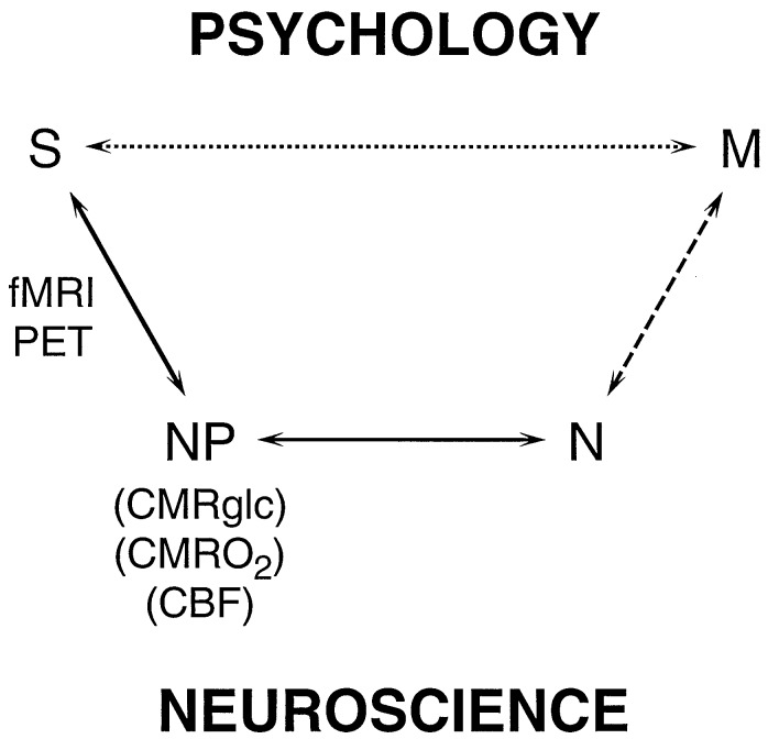
Figure 1.
Schematic relations between the signal (S) obtained in functional imaging experiments and mental processes (M). In the usual experimental plan and interpretation, based on psychology, a direct relationship between S and M is assumed, as represented by the upper pathway. The definition of M is based on psychology, while the imaging experiment serves to localize and quantitate the brain activity identified with the process. The lower pathway, Neuroscience, assumes that M has a molecular and cellular basis, which is broken into three steps leading to S. The signal, S, in fMRI or PET experiments, is primarily a measure of the neurophysiological parameters (NP) of cerebral metabolic rate of glucose consumption (CMRglc), cerebral metabolic rate of oxygen consumption (CMRO2) or CBF. PET methods have been developed for measuring each of these three parameters separately, while fMRI signals respond to differences in the changes of CBF and CMRO2, whose quantitative relationships are being investigated. CMRO2 and CMRglc measure cerebral energy consumption, while ΔCMRO2 and ΔCMRglc measure its increment. The relation between (NP) the neurophysiological measure of energy consumption and neuronal activity (N) has been clarified by the 13C MRS experiments (8–10). These recent findings allow measurements of S to be converted into measures of N, which places us squarely facing the unsolved “hard” problem of neuroscience, i.e., what is the relationship between M and N?

