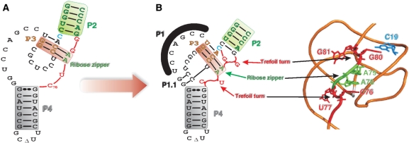
Figure 8.
Hypothetical representation of the folding of the J4/2 junction region before and after the formation of the P1.1 pseudoknot. (A) Nucleotide sequence and secondary structure of the antigenomic ribozyme characterized in this work. The substrate is represented by the thin line in order to simplify the representation. (B) 3D model representation of the antigenomic ribozyme drawn based on the backbone structure obtained from the crystal structure of the genomic version (3). The structural motifs are identified.

