Figure 1.
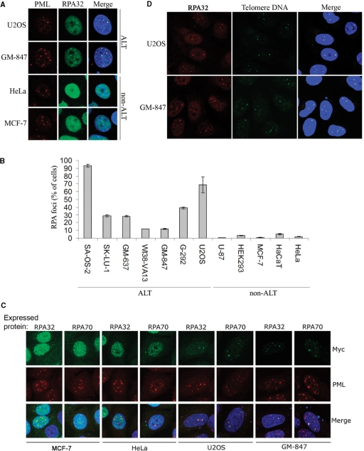
RPA foci that colocalize with PML-NBs are formed in ALT cells, but not in telomerase-positive cells. (A) HeLa, MCF-7, GM-847 and U2OS cells were grown on coverslips, fixed, and fluorescently labeled using antibodies against PML (red) and RPA32 (green). DAPI is shown in blue in the merged images. (B) Quantification of cells with RPA32-containing foci in ALT and non-ALT cell lines. Cells that contained one or more distinct RPA32 focus that colocalized with a PML-NB were scored. For each sample, between 600 and 800 cells were examined. Data represent two independent experiments ± standard deviation (SD). (C) Visualization of ectopically expressed myc-tagged RPA32 and RPA70 in ALT and non-ALT cell lines. Myc-tagged proteins and PML-NBs were fluorescently labeled using anti-myc (green) and anti-PML (red) antibodies, respectively. (D) Asynchronous GM-847 and U2OS cells grown on coverslips were subjected to FISH-IF using a FITC-conjugated telomeric PNA probe (green) and anti-RPA32 (red).

