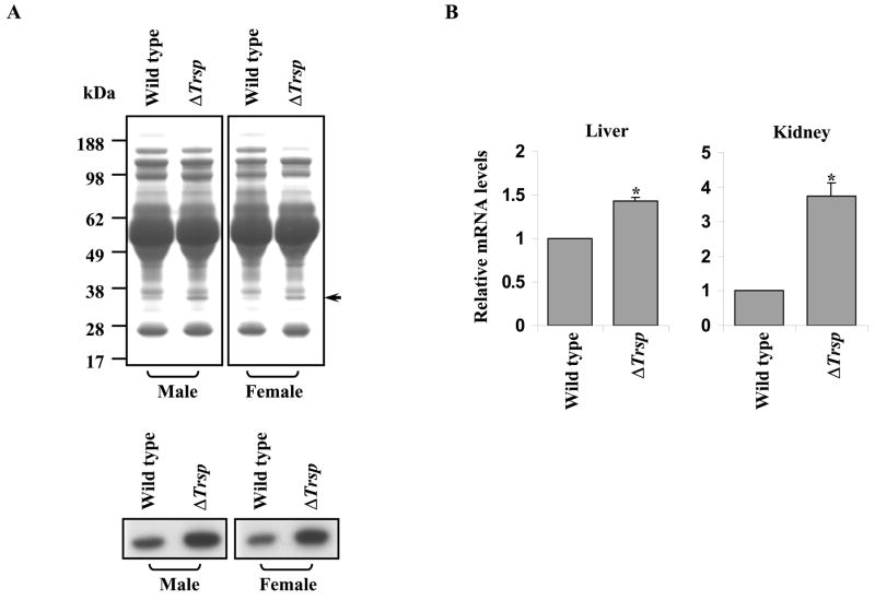
Fig. 2.
Analysis of ApoE and Apoe mRNA in wild type and ΔTrsp mice. (A) Coomassie Blue staining of plasma proteins from male and female wild type and ΔTrsp mice (upper panel) and immunodetection of ApoE using polyclonal anti-ApoE antibodies in plasma from the same mice (lower panel). (B) The relative Apoe mRNA levels in liver and kidney samples of wild type and ΔTrsp mice, determined by real-time PCR. The level of Apoe mRNA in each sample was normalized to that of 18S rRNA and the normalized value for Apoe mRNA in ΔTrsp mice was then plotted relative to that of wild type mice along with the error bars. The results are representations of 4 independent experiments, each carried out in triplicate (*, p < 0.0005 versus wild type mice).

