
Fig. 1. Photomicrographs of coronal sections through the dorsolateral (DL) DRN showing immunofluorescence for CRF and vGlut1.
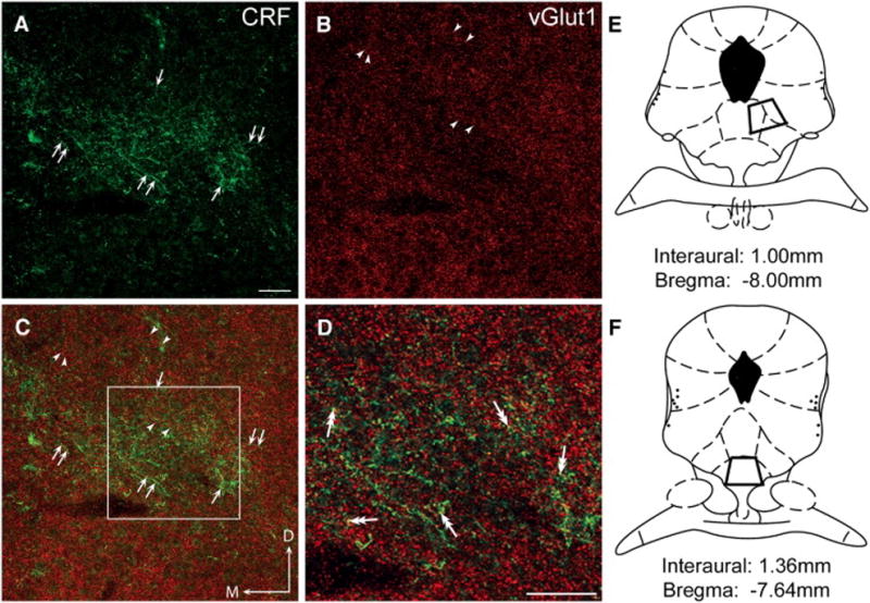
All images in these photomicrographs were obtained in the region of the DL DRN, ventral and to the right of the cerebral aqueduct, and dorsal to the medial longitudinal fasciculus. (A) CRF immunoreactivity was visualized using a FITC-conjugated secondary antibody (green) and the immunolabeled varicose fibers are indicated by arrows. The topographically organized fibers are consistent with previous reports of CRF-immunolabeled fiber distribution in this region and are also represented in panels C and D. (B) Vesicular glutamate transporter isoform1 (vGlut1) immunolabeling was detected using a TRITC-conjugated secondary antibody (red). Arrowheads indicate the punctate appearance of vGlut1 throughout the DL DRN in the same field as panel A. (C) The merged image illustrates the different patterns of distribution for both CRF (green; arrows) and vGlut1 (red; arrowheads) immunoreactivity in the DL DRN as depicted separately in panels A and B. Note that the CRF immunoreactivity is seen predominantly in varicose fibers, whereas vGlut1 is located in small puncta. (D) Dual-headed arrows indicate puncta that appear yellow in this higher magnification image of the region delineated by the square box in panel C, suggesting the presence of both CRF and vGlut1 in the same processes. (E and F) Regions sampled for both immunofluorescence and immunoelectron microscopic analysis of the DL DRN (E) and VM DRN (F) are illustrated in the schematic diagrams adapted from the Rat Brain Atlas [54] at level −8.00mm relative to bregma (DL DRN) and at level −7.64mm relative to bregma (VM DRN). Scale bar in A = 50 μm (also for B, C); scale bar in D = 50 μm. D:dorsal, M:medial