
Fig. 3. Ultrastructural evidence for the co-localization of CRF and vGlut1 within axon terminals of the DL DRN.
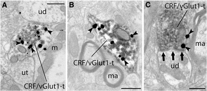
Silver enhanced gold particles (arrowheads) indicating CRF immunoreactivity were observed within axon terminals that contained immunoperoxidase associated with abundant clear synaptic vesicles indicating the presence of vGlut1 (A–D). (A) Although both unlabeled terminals (ut) and unlabeled dendrites (ud) could be identified throughout regions of the neuropil, individual terminals exhibited immunoreactivity for both CRF and vGlut1 (CRF/vGlut1-t). (B) As in all regions selected for analysis, immunoreactivity indicating the presence of CRF and vGlut1 was clearly localized to axon terminals, and here, myelinated axons (ma) lacking immunoreactivity can be seen in close proximity to the CRF/vGlut1-t. (C) Unlabeled dendrites (ud) were identified as one target of CRF/vGlut1-t, and here this CRF/vGlut1-t forms an asymmetric (excitatory) synapse (three grouped arrows) onto the unlabeled dendrite (ud). Scale bars=500nm.