Abstract
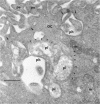
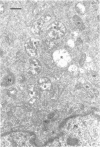
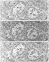
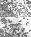
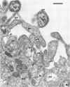
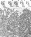
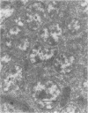
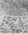
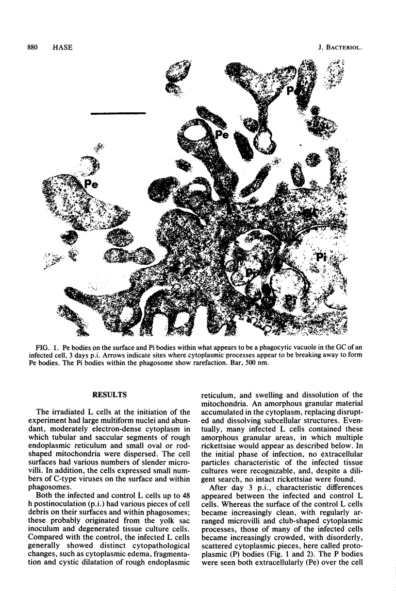
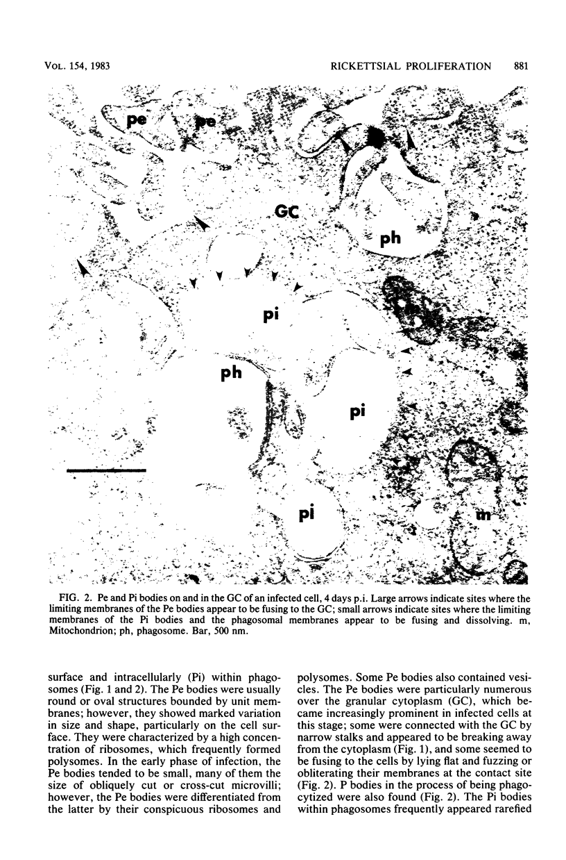
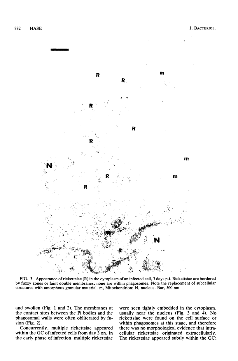
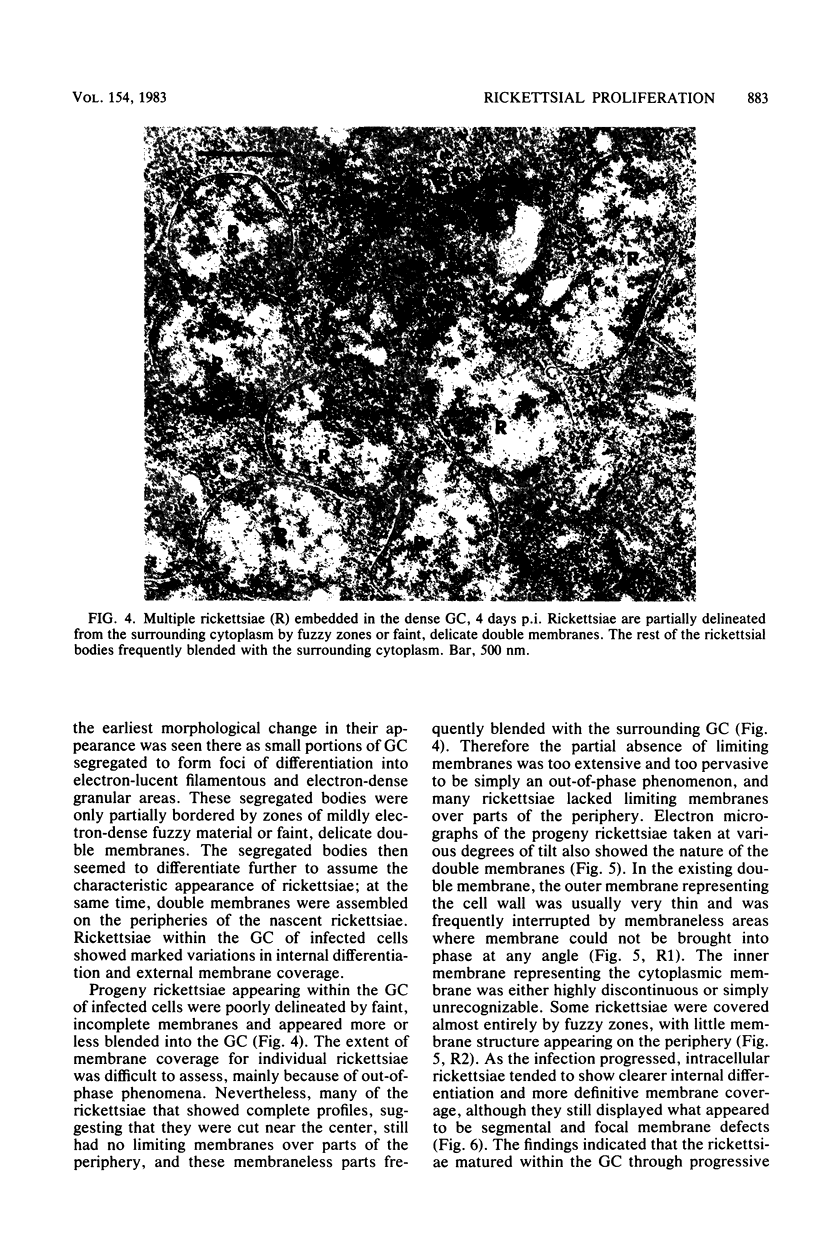
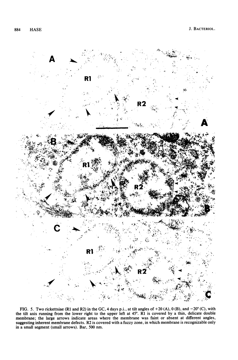
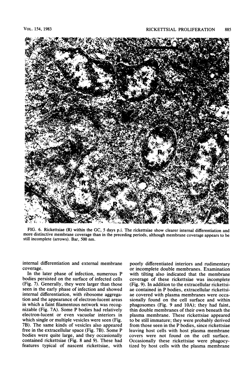
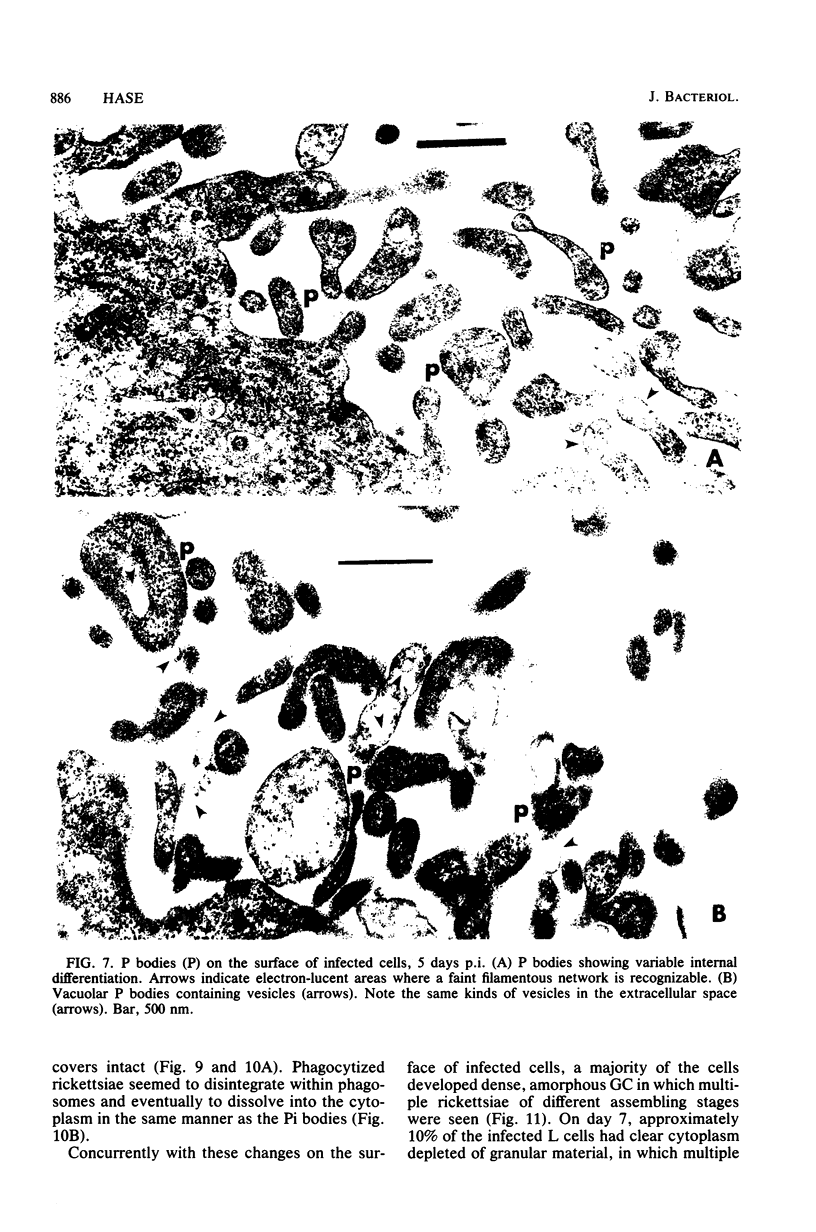
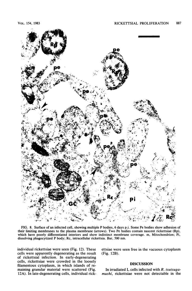
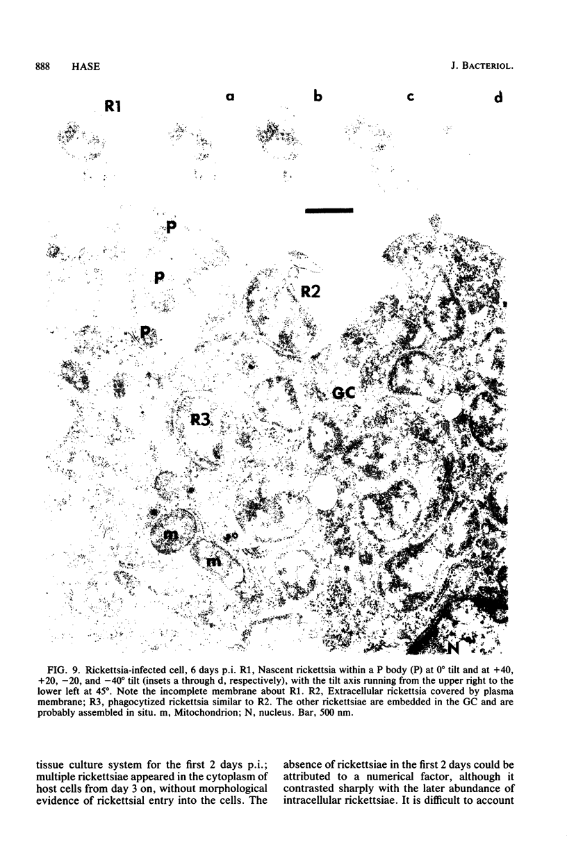
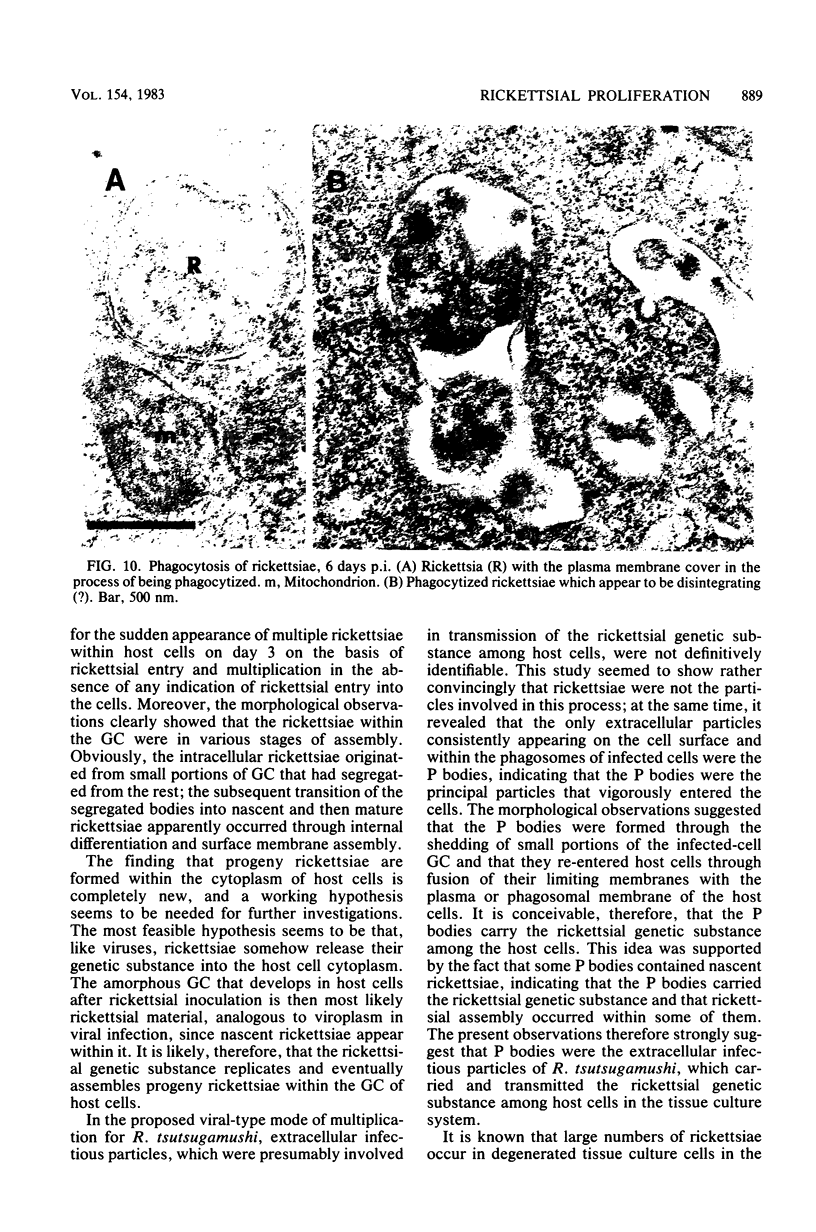
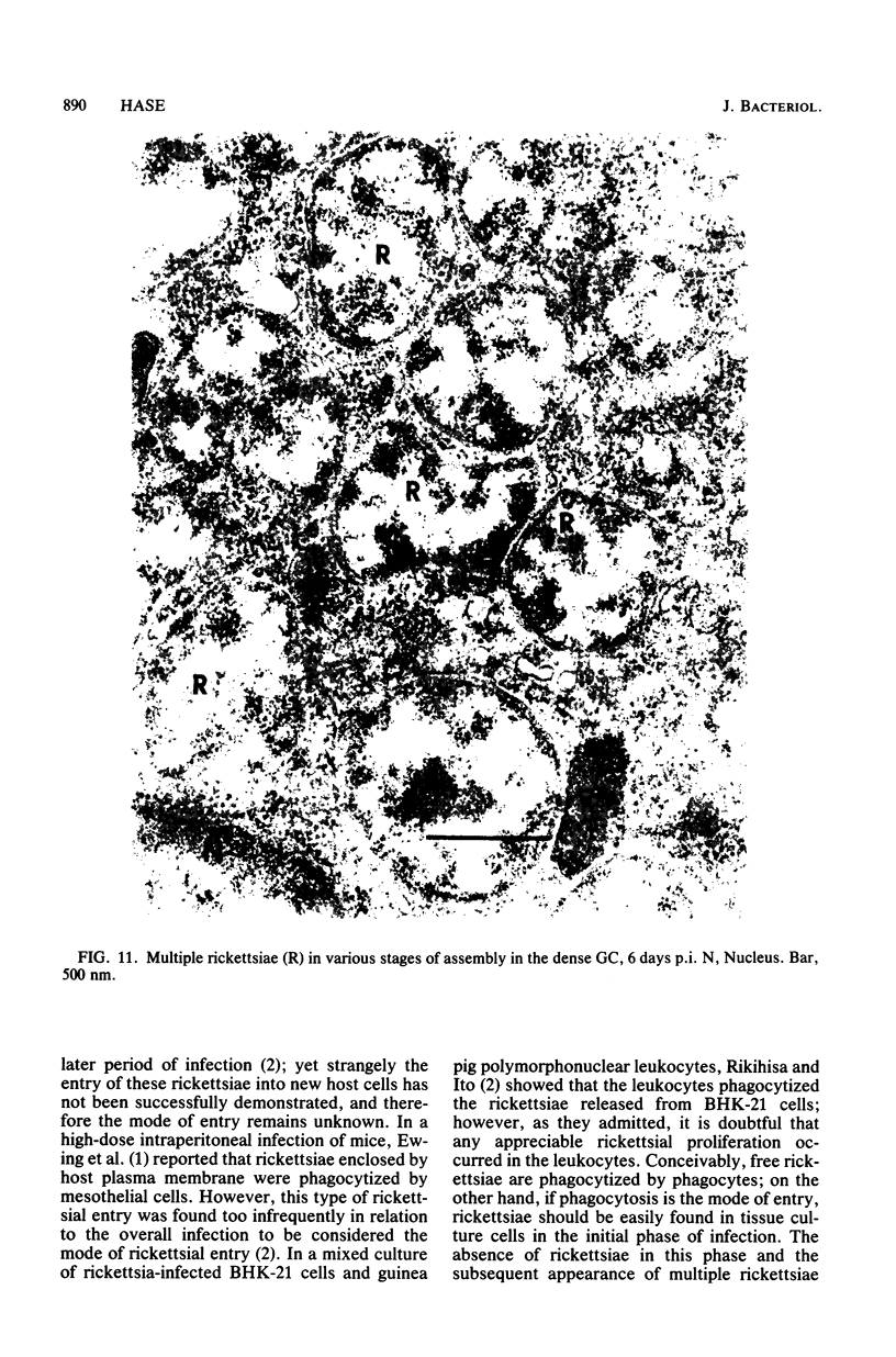
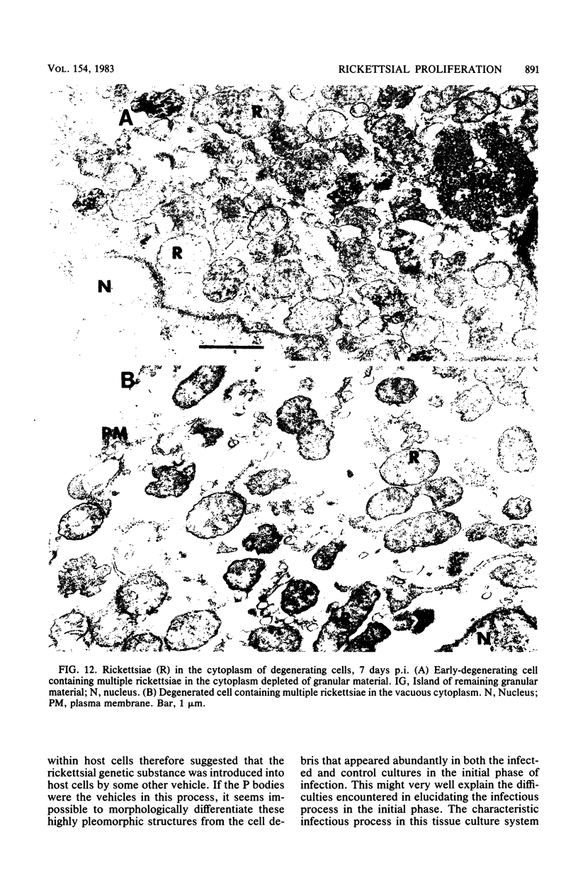
Irradiated L cells infected with Rickettsia tsutsugamushi were studied under the electron microscope to define the morphological growth pattern of the organism. For 2 days after inoculation, no rickettsiae were found either extra- or intracellularly; after 2 days multiple rickettsiae appeared within the host cells without morphological evidence of their entry. These observations showed that the rickettsiae within the cell were assembled in situ by segregation of portions of the granular cytoplasm and subsequent internal differentiation and surface membrane assembly of the segregated bodies. The protoplasmic (P) bodies, which seemed to be formed by shedding infected-cell granular cytoplasm, consistently appeared on the surface and within the phagosomes of the host cells. Rickettsiae were occasionally seen entering host cells in the later phase of infection; these were apparently the ones assembled within the P bodies. This suggested that the P bodies, and not the rickettsiae, were the major infectious particles that transmitted the rickettsial genetic substance among the host cells. On the basis of the present morphological observations, viral-type multiplication for R. tsutsugamushi is proposed.
Full text
PDF
Images in this article
Selected References
These references are in PubMed. This may not be the complete list of references from this article.
- Ewing E. P., Jr, Takeuchi A., Shirai A., Osterman J. V. Experimental infection of mouse peritoneal mesothelium with scrub typhus rickettsiae: an ultrastructural study. Infect Immun. 1978 Mar;19(3):1068–1075. doi: 10.1128/iai.19.3.1068-1075.1978. [DOI] [PMC free article] [PubMed] [Google Scholar]
- Rikihisa Y., Ito S. Localization of electron-dense tracers during entry of Rickettsia tsutsugamushi into polymorphonuclear leukocytes. Infect Immun. 1980 Oct;30(1):231–243. doi: 10.1128/iai.30.1.231-243.1980. [DOI] [PMC free article] [PubMed] [Google Scholar]
- Silverman D. J., Wisseman C. L., Jr Comparative ultrastructural study on the cell envelopes of Rickettsia prowazekii, Rickettsia rickettsii, and Rickettsia tsutsugamushi. Infect Immun. 1978 Sep;21(3):1020–1023. doi: 10.1128/iai.21.3.1020-1023.1978. [DOI] [PMC free article] [PubMed] [Google Scholar]
- Weiss E., Green A. E., Grays R., Newman L. M. Metabolism of Richettsia tsutsugamushi and Rickettsia rickettsi in irradiated host cells. Infect Immun. 1973 Jul;8(1):4–7. doi: 10.1128/iai.8.1.4-7.1973. [DOI] [PMC free article] [PubMed] [Google Scholar]


