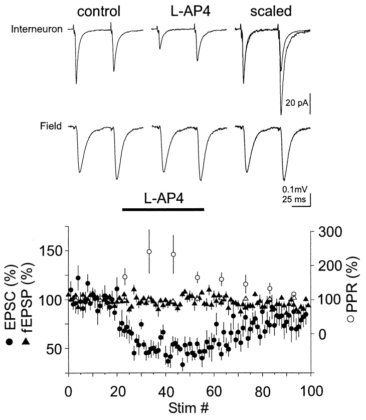
Figure 1.
L-AP4 reduces EPSCs recorded from interneurons in CA1 but not field EPSPs. Current and voltage traces: Data from a representative experiment in which field and whole-cell recordings were performed simultaneously. Each trace represents the average of 10–20 sweeps. (Lower) Summary graph of the action of L-AP4 (10 μM) on the amplitude of evoked AMPA receptor-mediated EPSCs recorded from CA1 interneurons voltage clamped at −70 mV (n = 11, •). Note the increase of the normalized PPR of the EPSCs recorded from interneurons upon perfusion of L-AP4 (○, bin size: 10 stimuli; the PPR was defined as (p2/p1) where p1 and p2 are the amplitude of the first and second EPSC, respectively). fEPSPs were monitored simultaneously by placing an extracellular-recording pipette in the stratum radiatum in six experiments (▴). No change in PPR could be detected in the field recordings (▵).

