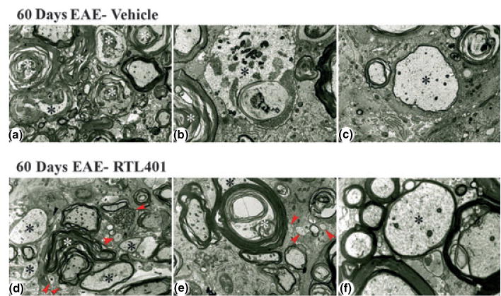
Fig. 7.

Representative electron micrographs showing lesion areas in spinal cords from mice with EAE on day 60 after treatment with vehicle (panels a–c) or RTL401 (panels d–f). (a) Low power view of typical lesion area showing marked continued Wallerian-like axonal degeneration (white asterisks) and demyelination (black asterisk). Note paucity of infiltrating cells and lack of small, regenerating axonal sprouts. Magnification × 4000. (b) Higher power view showing Wallerian-like axonal degeneration (white asterisk) and active demyelination (black asterisk). Magnification × 8000. (c) Higher power view of a large, remyelinating axon as shown by the relatively thin myelinated sheath (black asterisk). Magnification × 6700. (d) Low power view of typical lesion area showing continued Wallerian-like axonal degeneration (white asterisk), including a dystrophic axon (black arrow), and demyelinated and remyelinating axons (black asterisks). However, there are also prominent remyelinating axons and several small axonal sprouts (red arrowheads). Note paucity of infiltrating cells. Magnification × 4000. (e) Low power view of a large fiber (black asterisk) undergoing active demyelination (white asterisk). Note also three very small axons/regenerating sprouts (arrowheads). Magnification × 5000. (f) Higher power view of a medium-sized, remyelinating axon as shown by the relatively thin myelinated sheath (black asterisk). Magnification × 14 000.
