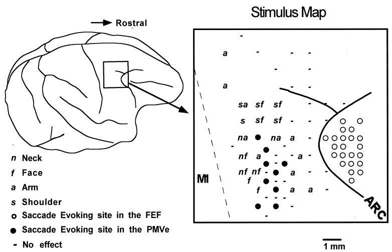
Figure 1.
(Left) Surface view of the cerebral cortex indicating the location of the frontal cortex penetrated with microelectrodes. (Right) Distribution of stimulus effects mapped on the cortical surface. Solid and open circles denote saccade-evoking sites in the PMV and FEF. Cortical sites evoking movements in the arm, face, neck, and shoulder are labeled a, f, n, and s, respectively. ICMS-negative sites are marked with −. The dashed line denotes the boundary between the primary and premotor cortex. ARC, arcuate sulcus.

