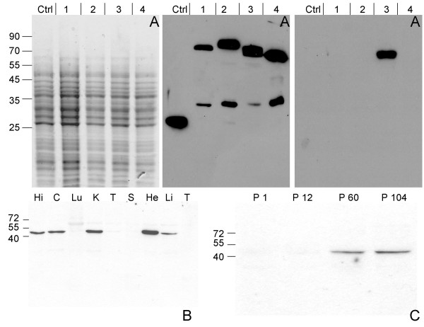
Figure 2.
Antibody specificity and Necl-3 expression. (Panel A) Crude lysates of S2 cells transfected with either GFP or one each of the Necl-GFP fusion proteins were visualized by SDS-PAGE and coomassie blue staining (left panel). The gels were blotted on nitrocellulose membranes and revealed with either an anti GFP mAb (middle panel), or the affinity-purified anti Necl-3 antibody (right panel). The cells had been transfected with GFP (Ctrl), Necl-1-GFP (1), Necl-2-GFP (2), Necl-3-GFP (3) or Necl-4-GFP (4). The anti GFP mAb reveals fusion proteins of the expected apparent molecular weights as well as faster migrating products, likely the result of degradation or internal translation initiation. The anti Necl-3 antibody does not cross-react with the other members of the Necl family. (Panel B) 15 μg of homogenates from hippocampus (Hi), cortex (C), lung (Lu), kidney (K), testis (T), spleen (S), heart (He), liver (Li) and thymus (T) of a 2-month-old rat were subjected to SDS-PAGE, followed by western blotting with the anti-Necl-3 antibody. (Panel C) Rat cortex homogenates from the indicated postnatal ages were subjected to SDS-PAGE (12%), followed by western blotting with the anti-Necl-3 antibody.

