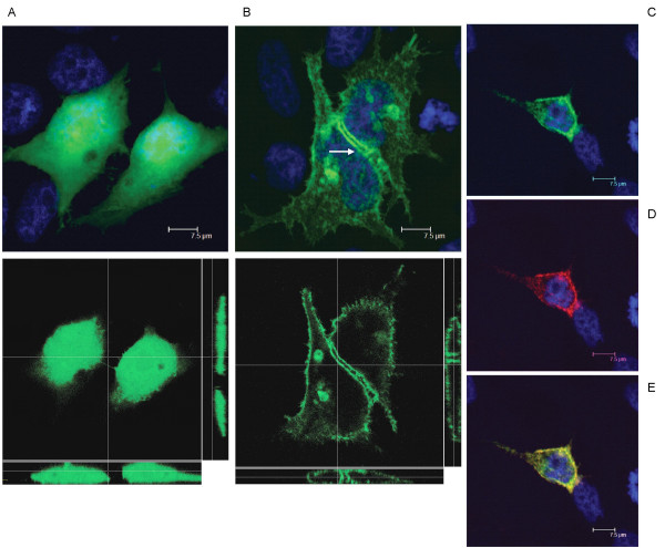
Figure 3.
GFP (A, top panel: average projection of 30 z-stacks of 0.24 micron each; bottom panel: a single X/Y plane in the center, a X/Z slice at bottom and a Y/Z slice on the right) or Necl-3-GFP (B, top panel: average projection of 39 z-stacks of 0.12 micron each; bottom panel: a single X/Y plane in the center, a X/Z slice at bottom and a Y/Z slice on the right) was transfected in Hela cells and visualized by confocal fluorescence microscopy. Unlike GFP alone, Necl-3 accumulates in the plasma membrane and at cell-cell contacts between transfected cells (arrow). (C-E), Hela cells transfected with Necl-3-GFP were processed for immunofluorescence with the anti Necl-3 antibody in non-permeabilized conditions and visualized (average projection of 83 z-stacks of 0.12 micron each) in the GFP channel (C) and in the rhodamine channel (D). To reveal untransfected cells, the specimen was stained with DAPI. The (E) panel shows the merged DAPI, GFP and rhodamine channels. Note that the Necl-3 antibody only stains the transfected cells.

