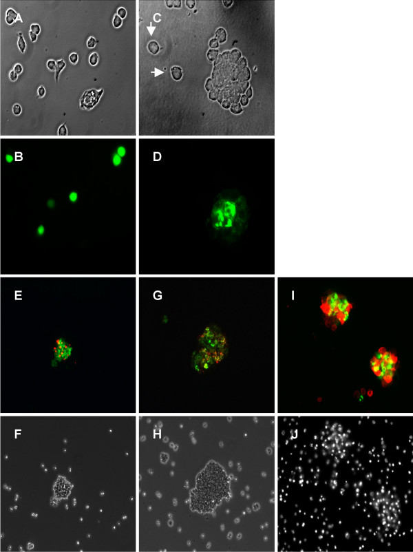
Figure 5.
S2 cells transfected with GFP (A, B) or Necl-3-GFP (C, D) were gently shaken and visualized by phase contrast (A, C) and fluorescence microscopy (B, D). Cell aggregates form with Necl-3-GFP transfected cells (C, D), whereas GFP transfected (A, B) and untransfected cells (arrows in C) do no aggregate. S2 cells transfected with Necl-3-GFP were mixed with Necl-1-DsRed cells (E, F); cells transfected with Necl-3-GFP were mixed with Necl-4-DsRed (G, H) cells, and cells transfected with Necl-3-myc were mixed with Necl-2-GFP (I, J). The cell aggregates were visualized by fluorescence microscopy with green and red pass filters (E, G, I), by phase contrast (F, H), or by fluorescent staining of the nuclei with DAPI (J).

