Fig. 3.

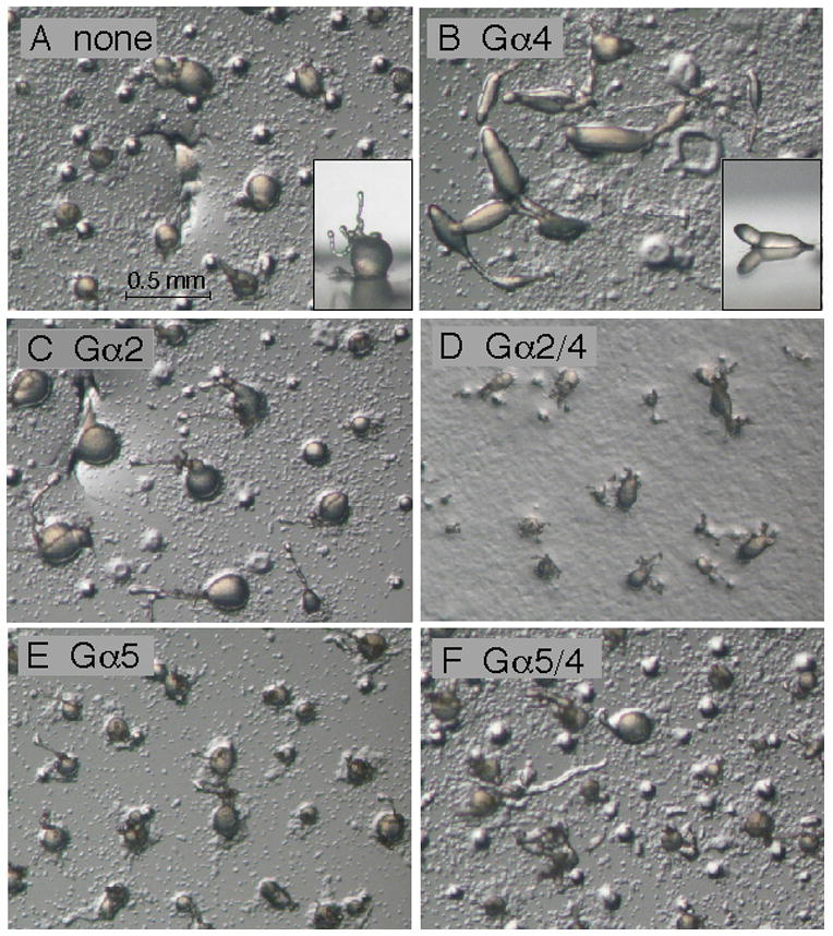
Development of gα4− cells with or without Gα subunit expression vectors. Cells were grown in axenic medium and washed in phosphate buffer and then cell suspensions (2 × 10⁸ cells/ml) were plated for development as described in the Materials and methods section. Panel (A) aggregates of gα4− cells with no Gα subunit expression vector. Insert displays a side view of a typical aggregate with the round mound and extended tip morphology. Panel (B) aggregates of gα4− cells with Gα4 subunit expression vector in the early stages of culmination. Insert displays a side view of a typical aggregate displaying wild-type slug morphology. Panel (C) gα4− cells with Gα2 subunit expression vector. Panel (D) gα4−cells with Gα2/4 subunit expression vector. Panel E: gα4− cells with Gα5 subunit expression vector. Panel F: gα4− cells with Gα5/4 subunit expression vector. Developing cell aggregates were photographed 23 hrs after initial plating using a dissecting microscope (20X magnification).
