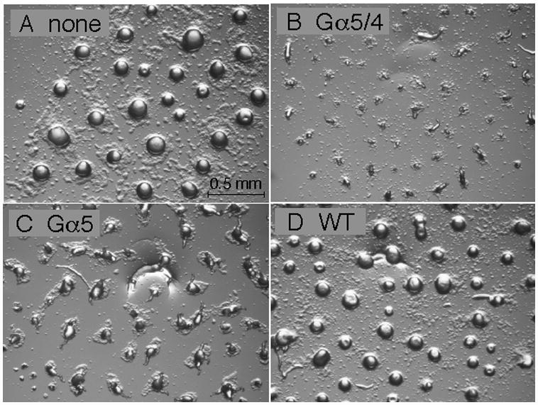
Fig. 5.

Development of gα5− cells with or without Gα subunit expression vectors. Cells were grown in axenic medium and washed in phosphate buffer and then cell suspensions (2 × 10⁸ cells/ml) were plated for development as described in the Materials and methods section. Panel (A) aggregates of gα5− cells with no Gα subunit expression vector. Panel (B) aggregates of gα5− cells with Gα5/4 subunit expression vector. Panel (C) aggregates of gα5− cells with Gα5 subunit expression vector. Panel (D) aggregates of wild-type cells with no Gα subunit expression vector. Developing cell aggregates were photographed 12 hrs after initial plating using a dissecting microscope (20X magnification).
