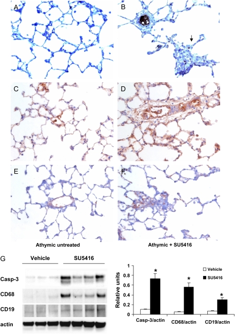
Figure 3.
Immunohistochemical staining of activated caspase-3 (A, B); CD68 (C, D), and CD20 (E, F) of untreated (A, C, E) and SU5416-treated (B, D, F) athymic rat lungs. Original magnification, ×400. (G) Western blot and quantitative analysis of activated caspase-3 (2 wk after SU5416 injection), CD68, and CD19 (3 wk after SU5416 injection) in the whole lung extracts of untreated and SU5416-treated athymic rats. *p < 0.01.

