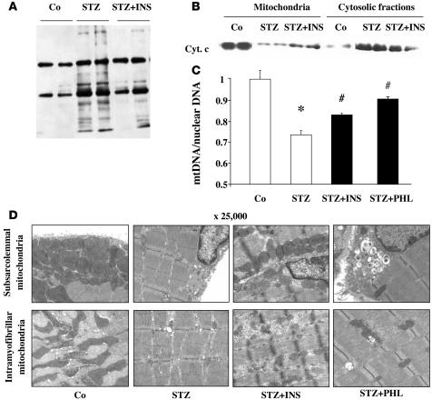
Figure 5. STZ-induced oxidative stress alters mitochondria density and structure in skeletal muscle.
(A) Immunoblots showing total protein carbonylation in the gastrocnemius muscle of control (Co), STZ, and insulin-treated STZ (STZ+INS) mice. (B) Immunoblot showing cytochrome c protein in the MF and CF of the gastrocnemius muscle of control, STZ, and insulin-treated STZ mice. (C) mtDNA copy number was calculated as the ratio of COX1 to cyclophilin A DNA levels, determined by real-time PCR, in the skeletal muscle of control, STZ, insulin-treatd STZ, and phlorizin-treated STZ (STZ+PHL) mice (n = 6). Note that the y axis scale is between 0.5 and 1. Results were normalized by the mean value for the control mice set to 1 unit. *P < 0.01 vs. control; #P < 0.05 vs. STZ. (D) Transmission electron microscopy images (original magnification, ×25,000) of subsarcolemmal and intermyofibrillar mitochondria from the gastrocnemius muscle of control, STZ, insulin-treated STZ, and phlorizin-treated STZ mice.

