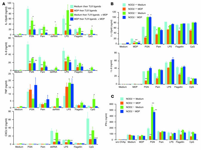
Figure 6. Human and mouse DCs prestimulated with MDP exhibit reduced cytokine and chemokine production when stimulated with TLR ligands.
(A) Human monocyte–derived DCs (1 × 10⁶/ml) from 6 healthy donors were preincubated with MDP or medium for 24 hours and then stimulated with a broad range of TLR ligands alone or in combination with MDP for an additional 24 hours. Cultured supernatants were collected at 24 hours and analyzed for cytokine and chemokine production by ELISA. *P < 0.05; **P < 0.01 compared with supernatants from DCs preincubated with medium and stimulated with TLR ligands alone (light blue bars). (B) CD11c+ DCs (1 × 10⁶/ml) derived from bone marrow cells from NOD2-intact (NOD2+/+) and NOD2-deficient (NOD2–/–) mice were preincubated with MDP (50 μg/ml) or medium alone for 24 hours and stimulated with a broad range of TLR ligands. Cultured supernatants were collected at 24 hours and analyzed for cytokine production by ELISA. *P < 0.05; **P < 0.01 when supernatants were compared with NOD2-intact DCs preincubated with medium and stimulated with TLR ligands (light blue bars). (C) OVA323-339 peptide–specific CD4+ T cells (OT-II) were purified from the spleens of OT-II transgenic mice; OT-II cells (1 × 10⁶/ml) were cocultured with NOD2-intact or NOD2-deficient BMDCs (2 × 10⁶/ml) in the presence of a broad range of TLR ligands and OVA peptide (0.5 μM); cultured supernatants were collected at 72 hours and analyzed for IFN-γ production by ELISA. *P < 0.05; **P < 0.01 compared with supernatants from NOD2-intact DCs preincubated with medium and stimulated with TLR ligands (light blue bars).

