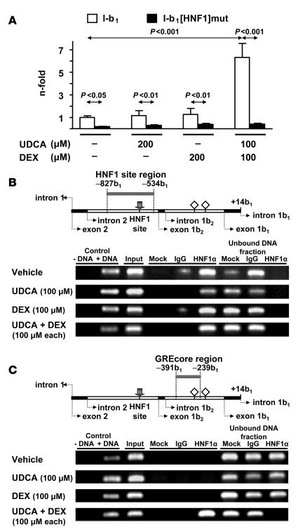
Figure 11. Role of the HNF1α and its interaction with the HNF1 element for the effects on AE2 alternate promoter following simultaneous treatment of PLC/PRF/5 cells with UDCA and dexamethasone.
(A) Luciferase activities of PLC/PRF/5 cells transiently transfected with wild-type construct I-b1 or with HNF1-mutated construct I-b1[HNF1]mut and treated for 24 hours with UDCA and/or dexamethasone. Values of luciferase activity (normalized with Renilla values) are given as fold activity relative to the activity in cells transfected with construct I-b1 treated with just vehicle (i.e., in the absence of UDCA and DEX). Data are mean ± SD; n = 9 each. (B) ChIP assays with HNF1α immunoprecipitates from extracts of PLC/PRF/5 cells treated as described in Figure 10, in which the region that includes the HNF1 element was amplified (amplicon –827b1/–534b1; see the gray bar in the upper diagram). Positive and negative controls were equivalent to those in Figure 10A, but IgG for the negative immunoprecipitation control was goat total IgG. (C) ChIP assays with HNF1α immunoprecipitates in which the region that includes GREcore –327b1 (amplicon –391b1/–239b1 indicated by a gray bar in the upper diagram) was amplified using the same templates as in B. In upper diagrams of B and C, gray arrows indicate the HNF1 site; open rhombuses represent GREcore motifs.

