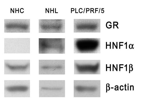
Figure 13. Western blot analysis using whole-cell extracts from NHCs and PLC/PRF/5 cells.
Total lysate from normal human liver (NHL) (shown in between) was used for a comparison with a normal expression pattern of these proteins in whole liver tissue. Electrophoresed proteins were electrotransferred and probed with antibodies against GR, HNF1α, and HNF1β. Loading control was carried out with an antibody against β-actin.

