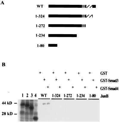
Figure 2.
In vitro association of Smad3 and Smad4 with JunB. (A) Schematic diagram of JunB deletion mutants used in the binding study shown in B. (B) In vitro association of JunB and JunB deletion mutant TNT products with GST, GST-Smad3, or GST-Smad4. Bacterially produced GST, GST-Smad3, and GST-Smad4 proteins were coupled to glutathione-Sepharose, incubated with the indicated TNT product, centrifuged, and the Sepharose-bound proteins visualized by Coomassie stained SDS/PAGE (data not shown). The amount of each GST protein and the volume of glutathione-Sepharose used in each binding reaction were normalized. Each reticulocyte lysate was produced as described in Materials and Methods. Domains depicted in A refer to the same domains depicted in Fig. 1. Five percent of each reticulocyte lysate input was run in lanes 1–4: lane 1, JunB full length; lane 2, 1–324; lane 3, 1–272; lane 4, 1–80.

