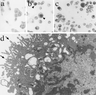
Figure 2.
Characterization of the signet ring-like cells formed by the expression of pBD110 in HCC2998/BD110 cells. (a and b) PAS staining of mucinous substance in the pBD110-expressing cells (b) or control cells (a). HCC2998/BD110 cells were cultured on a coverslip for 3 days after infection with AxCANCre, fixed with 4% paraformaldehyde, and PAS stained. Eccentric nuclei and large droplets containing diastase-resistant PAS-positive substance can be seen in the signet ring-like cells (indicated by an arrowheads). (c) Intracellular localization of a secretory glycoprotein antigen, CA15–3. Cells prepared as above were embedded in paraffin and the section was stained with anti-CA15–3 antibody. CA15–3 antigen was detected in various sized vacuoles in cytoplasm as well as the entire cell surface. Various cross sections of the cells were observed. (d) Electron micrograph of a signet ring-like cell found in the BD110-expressing cells. HCC2998/BD110 cells cultured for 3 days after AxCANCre infection were used to prepare the section. A typical signet ring-like cell was photographed. Dilation of the Golgi apparatus was observed from perinuclear to the submembranous regions (indicated by arrowheads). Irregular elongation of microvilli was localized above the dilated Golgi apparatus (indicated by arrows). (Original magnification, ×4000.)

