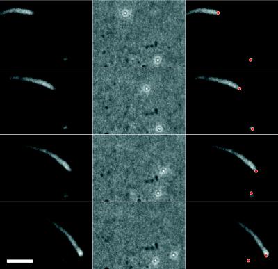
Figure 1.
Actin-based movement of a 0.5-μm-diameter carboxylated polystyrene microsphere uniformly coated with ActA-His. ActA-His-coated microspheres were incubated in Xenopus egg cytoplasmic extract supplemented with 0.15 mg/ml rhodamine-actin. Four frames from a video sequence are shown, each separated by 30 sec. A stationary bead surrounded by an actin cloud is at the lower right. (Left column) Fluorescence image showing distribution of rhodamine-actin in the comet tail and cloud. (Center column) Phase-contrast image showing position of polystyrene beads. (Right column) Bead positions (red dots) superimposed on fluorescence images. This bead is moving at a rate of 0.153 μm/sec. (Bar = 5 μm.)

