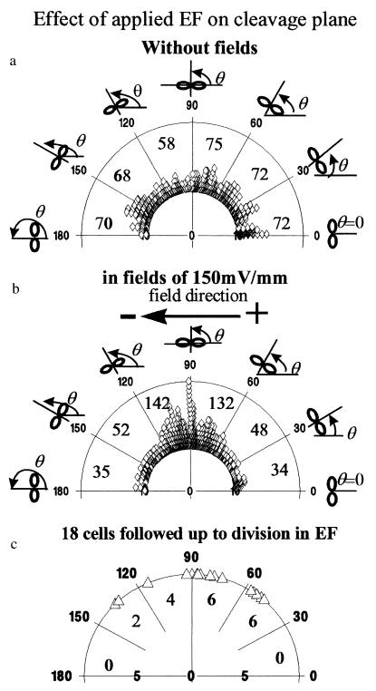
Figure 3.
Angles of cleavage plane of transformed human corneal epithelial cells expressed as a polar diagram, where each symbol represents one cell. (a) Angles of cleavage plane of 415 dividing cells cultured without electric fields. (b) Polarized orientation of the cleavage plane of 443 cells dividing in a small physiological EF (in EFs for 20 hr in 5% CO2 incubator/37°C). Clearly, a high proportion of cells divided with a cleavage plane orthogonal to the EFs. (c) Polarized cleavage plane orientation of 18 dividing cells followed continuously in EF on the microscope stage. The total number of cells in each range of angles is indicated, both numerically and by compression of the symbols.

