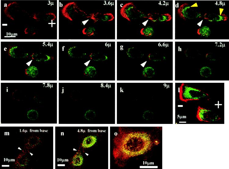
Figure 4.
(a–k) Confocal optical sections of a dividing bovine corneal epithelial cell in an applied EF (at 150 mV/mm in DMEM/10% FBS). F-actin is concentrated at the cleavage plane (white arrowhead). Additionally, there is prominent accumulation of F-actin (red) and of TGFR II (green) at either poles of the two daughter cells (especially b–e). Although this segregation of F-actin and TGFR II to the two poles is evident, a nondividing cell beneath and two other nondividing cells (l) showed predominant accumulation of F-actin and TGFR II on the cathodal side. (m–o) Dividing cells (m and n) and nondividing cells (o) cultured without an applied EF are shown. Staining in both cases is more diffuse than for EF-treated cells, and, with the exception of some F-actin accumulation in the cleavage furrow, there was little polarization of F-actin or of TGFR II.

