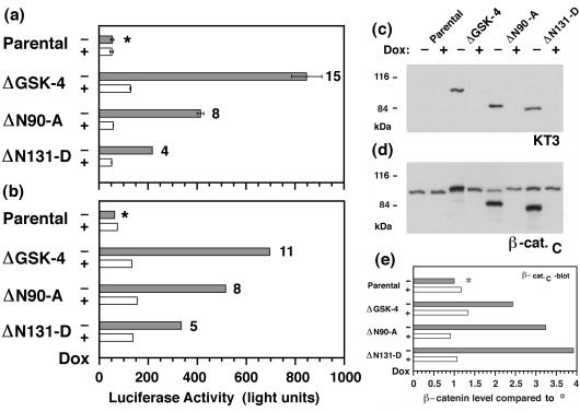
Figure 3.
Increased TCF reporter activity in MDCK clones expressing mutant β-catenin. (a and b) Parental MDCK cells and MDCK clones expressing mutant β-catenin were preincubated for 3 days without or with Dox (−/+Dox) and then cotransfected with pTOPFLASH/pSV-β-galactosidase. Luciferase activities (relative light units) were corrected for differences in transfection efficiencies, which were estimated by β-galactosidase activities in the same samples. Data from two independent experiments are summarized in a and b; bars represent mean values from two independent samples (a) or one sample (b). Numbers at the head of the bars represent “fold activation” compared with luciferase activity in parental cultures without (−) Dox (∗) for each experiment. TCF-activated transcription of the luciferase reporter was higher in clones expressing the mutant β-catenins (−Dox, shaded bars) compared with cultures in which expression of mutant β-catenins was repressed (+Dox, open bars) or with parental cells. (c and d) Parallel cultures to those reported in a were extracted with 1% SDS, and 15-μg protein lysates were subjected to SDS/PAGE and immunoblotted with mAb KT3 (c) or mAb β-cat.C (d). Molecular mass standards are indicated in kDa. (e) Levels of total (sum of endogenous and mutant) β-catenin were quantified from the β-cat.C immunoblot. x axis represents “fold expression” compared with the level of endogenous β-catenin in the parental culture/−Dox (∗).

