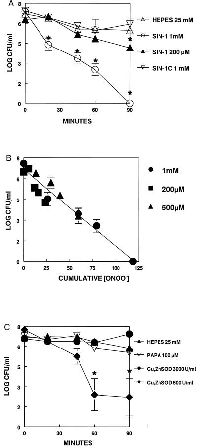
Figure 5.
M. pulmonis was grown to late logarithmic phase, washed to remove serum, and resuspended in 10 ml of 25 mM Hepes buffer, pH 7.4. Aliquots were taken at 0, 20, 45, 60, and 90 min for determination of cfu. (A) Hepes 25 mM, mycoplasmas alone; SIN-1 1 mM, mycoplasmas + 1 mM SIN-1; SIN-1 200 μM, mycoplasmas + 200 μM SIN-1; SIN-1C, mycoplasmas + 1 mM SIN-1C. (B) cfu vs. ONOO− concentration for the indicated concentrations of SIN-1 as measured by dihydrorhodamine 123 oxidation. (C) Hepes 25 mM, mycoplasmas alone; PAPA 100 μM, mycoplasmas +100 μM PAPANONOate; Cu, ZnSOD 3,000 units/ml, mycoplasmas +1 mM SIN-1 + 3,000 units/ml Cu, ZnSOD.;Cu, ZnSOD 500 units/ml, mycoplasmas +1 mM SIN-1 + 500 units/ml Cu, ZnSOD. ∗, significant difference between control and experimental groups at each time point, P <0.05.

