
Figure 7.
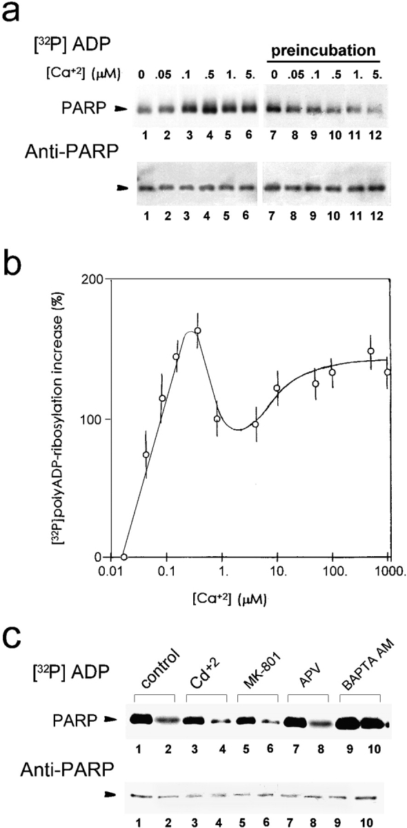
Extra-nuclear Ca2+ promotes polyADP-ribosylation of PARP. (a) PolyADP-ribosylation of PARP in crude nuclei isolated from unstimulated neurons, exposed to increasing [Ca2+], 1 min before (preincubation) or during [32P]polyADP-ribosylation (2 min, 25°C). [32P]polyADP-ribosylated PARP was immunoprecipitated, electroblotted (Western blot), autoradiographed, and immunolabeled by Vic-5 antibody. (b) The curve shows the average increase (±SD) in [32P]polyADP-ribosylation of PARP (measured by densitometry) versus the increase in extranuclear [Ca2+], relative to PARP [32P]polyADP-ribosylation in extranuclear [Ca2+] = 25 nM (see Materials and Methods) (n = 9). (c) Back-[32P]polyADP-ribosylation of PARP, in nuclei of depolarized or unstimulated neurons treated with Ca2+ influx blockers and the permeant chelator BAPTA AM. (Top) Autoradiograms of [32P]polyADP-ribosylated PARP in crude nuclei of unstimulated neurons (lanes 1, 3, 5, 7, and 9), and neurons depolarized by a 10-min train of repetitive (10 Hz) 30-volt, 0.1 ms pulses (lanes 2, 4, 6, 8, and 10). Cultured neurons were preincubated for 10 min at 25°C with 1 mM CdCl2 (lanes 3 and 4) or with antagonists of NMDA-glutamate receptors, MK-80l (20 μM; lanes 5 and 6) and APV (500 μM; lanes 7 and 8), or preincubated (30 min, 25°C) with the permeant Ca2+ chelator, BAPTA AM (50 μM; lanes 9 and 10). PARP was immunoprecipitated from the nuclear protein extracts of these neurons by N-20 antibody, subjected to SDS-PAGE, electroblotted (Western blot), and autoradiographed. (Bottom) Immunolabeling of [32P]-polyADP-ribosylated PARP by Vic-5 antibody in the immunoprecipitates (n = 6).