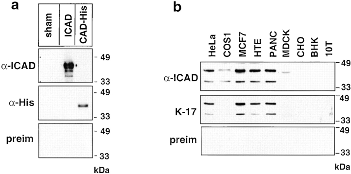
Figure 1.
Characterization of the anti-hICAD antibody. (a) The polyclonal rabbit anti-ICAD immune serum recognizes recombinant hICAD on immunoblot. Full-length hICAD and mCAD-(His)6 were expressed in BL21(D3) cells, and the bacterial lysates (∼10 μg protein/lanes) were separated with SDS-PAGE, transferred to nitrocellulose, and polypeptides were visualized by enhanced chemiluminescence using rabbit polyclonal anti-hICAD antiserum (α-ICAD), mouse monoclonal anti-His (α-His) primary antibody, or preimmune serum with the corresponding HRP-conjugated secondary antibody. Lysate obtained from sham-transformed bacteria were used as negative controls. (b) Western blot analysis of endogenous hICAD expression. Equal amounts of protein (50 μg) of the indicated cell lysate were separated with SDS-PAGE and subjected to immunoblotting as described in Materials and Methods. Two prominent immunoreactive polypeptides, with an apparent molecular mass of ∼45 and ∼36 kD, corresponding to the full-length ICAD (ICAD-L) and the alternatively spliced variant ICAD-S, respectively, are recognized with the rabbit (α-ICAD) and goat (K-17) anti-hICAD antibodies but not with the rabbit preimmune serum (preim).

