Figure 7.

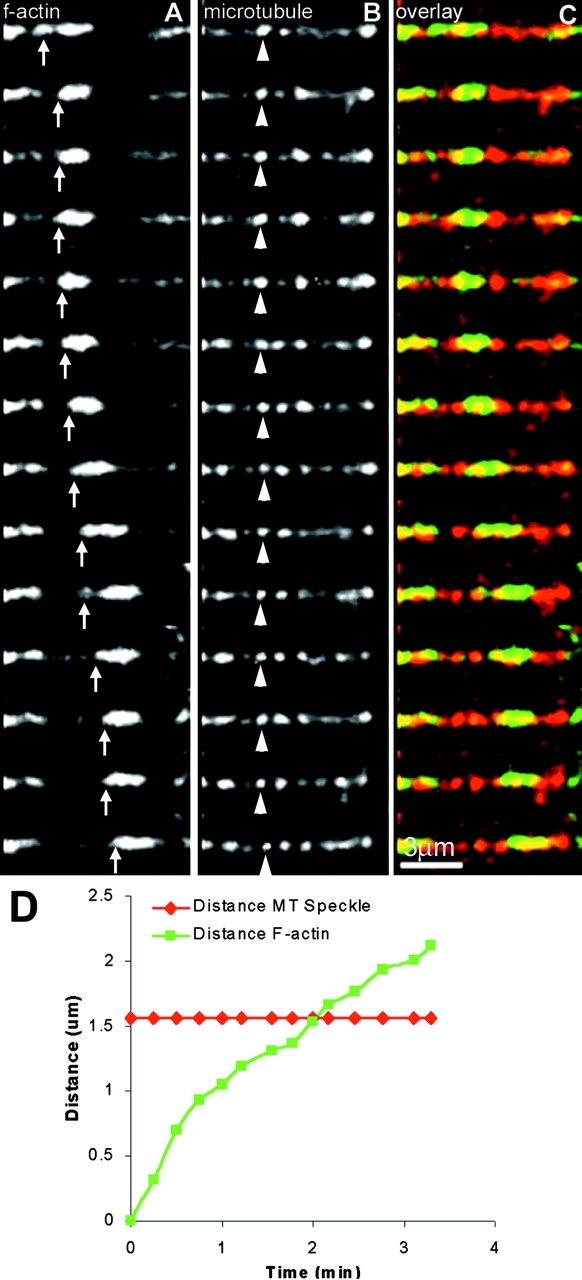
F-actin can translocate along the lattice of stationary microtubules. (A) Time-lapse fluorescence imaging shows F-actin moving in a straight path. (B) Time-lapse FSM view of a stationary microtubule in the same field of view shown in A. The speckle mark indicated by the arrowhead does not move with respect to the substrate, showing that this microtubule is stationary throughout the period of imaging. (C) Color overlay of F-actin from A and the microtubule from B shows that the F-actin moves with respect to the stationary speckle marks on the microtubule lattice. Images in A–C were acquired at 15-s intervals (also see supplemental video 12 at http://www.jcb.org/cgi/content/full/150/2/361/DC1). (D) Plot of the distance of the tip (arrow in A) of the F-actin bundle and the speckle mark on the microtubule (arrowhead in B) from the origin (the position of the F-actin bundle tip at time 0:00) vs. time. This demonstrates that the F-actin moves relative to the microtubule lattice.
