
Figure 4. DNA is fragmented and caspases are active when salivary gland growth is maintained.
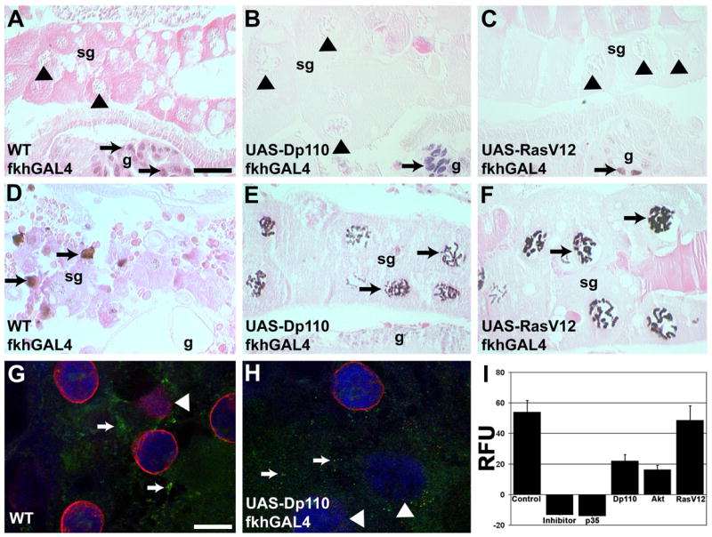
(A–F) Visualization of DNA fragmentation by the TUNEL assay. Nuclei of 6-hour apf (A) Canton-S control, (B) Dp110-, and (C) RasV12-expressing salivary glands are TUNEL negative (black triangles) while nuclei in nearby dying gut cells are TUNEL positive (black arrows). TUNEL positive nuclei were detected in salivary glands from 13.5-hour (D) Canton-S control, (E) Dp110- and (F) RasV12-expressing salivary glands. (G, H) Antibodies against processed caspase-3 (green) and nuclear lamins (red) indicate that processed caspase-3 is present (white arrows) and that the caspase substrate lamin is degrading (white arrowheads) in 13.5-hour (G) wild-type control glands and (H) Dp110-expressing glands (DNA is blue). (I) Cleavage of the caspase substrate Z-DEVD-AMC was measured in 13.5-hour salivary glands of Control Canton-S, Control Canton-S plus Ac-DEVD-CHO as an inhibitor, and of p35-, Dp110-, Akt- and RasV12-expressing salivary glands. Scale bar in (A) is 50 μm and (A–F) are the same magnification. Scale bar in (G) is 30 μm and (G–H) are the same magnification. Symbols are (sg) salivary gland, (g) gut, and (RFU) relative fluorescence units.