
Figure 5. Multiple degradation pathways are required for complete salivary gland histolysis.
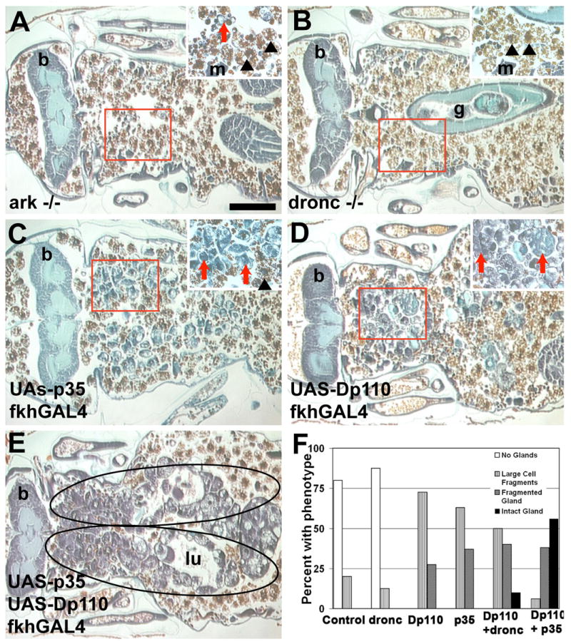
(A–E) Paraffin sections 24 hours apf. Salivary glands are degraded in (A) arkL46/N28 and (B) droncI24/I29 mutants that develop normally. Partial degradation occurs in salivary glands expressing (C) p35 and (D) Dp110. (E) Expression of p35 combined with Dp110 significantly increases the amount of persistent salivary gland tissue (encircled). (F) Histological sections of the indicated genotypes were evaluated for the amount and type of salivary gland tissue that was present in pupae 24 hours apf. The percentage of pupae with each phenotype is presented (n > 20 pupae/genotype). Scale bar in (A) is 200 μm and (A–E) are the same magnification. Red boxes indicate the region of higher magnification shown in the insets for each image. Red arrows point to salivary gland fragments. Black arrowheads point to fat body that is present in the area after the salivary gland has degraded. Symbols are (b) brain, (g) gut, (m) muscle, and (lu) lumen.