
Figure 6. Loss-of-function autophagy mutants inhibit salivary gland cell degradation in the presence of caspases.
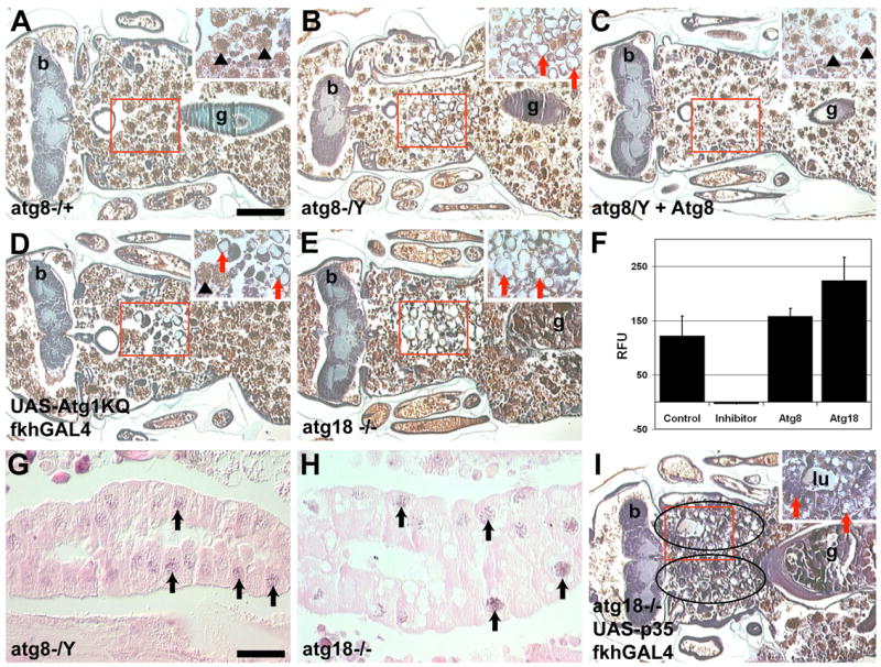
(A–E) Histological sections 24 hours apf. (A) Salivary glands are degraded in atg8aKG07569/Canton-S control pupae. (B) atg8aKG07569 mutant pupae contain vacuolated salivary gland cellular fragments. (C) Heat shock induced expression of hsdAtg8a-GFP rescued the vacuolated salivary gland phenotype of atg8aKG07569 mutant pupae. (D) Atg1KQ expression prevents complete salivary gland destruction and vacuolated salivary gland cell fragments persist. (E) atg18KG03090/Df(3L)66C-G28 mutant pupae contain vacuolated salivary gland cell fragments. (F) Cleavage of the caspase substrate DEVD-AMC was measured in whole pupae at 4 hours apf in: Canton-S control, Canton-S plus Ac-DEVD-CHO inhibitor, atg8aKG07569 mutant pupae and atg18KG03090/Df(3L)Exel6112 mutant pupae. Data is presented as the mean ± SE. (G–H) TUNEL positive nuclei (black arrows) are present in salivary glands of (G) atg8aKG07569 and (H) atg18KG03090/Df(3L)Exel6112 mutant pupae. (I) Expression of p35 in the salivary glands of atg18KG03090/Df(3L)Exel6112 mutant pupae leads to increased salivary gland persistence. Scale bar in (A) is 200 μm and (A–E and I) are the same magnification. Scale bar in (G) is 50 μm and (G–H) are the same magnification. Red boxes indicate the region of higher magnification shown in the insets for each image. Red arrows point to salivary gland fragments. Black arrowheads point to fat body that is present in the area after the salivary gland has degraded. Symbols are (b) brain, (g) gut and (lu) lumen.