
Fig. 2. Global and local neocortical DOWN states (delta waves) during SWS.
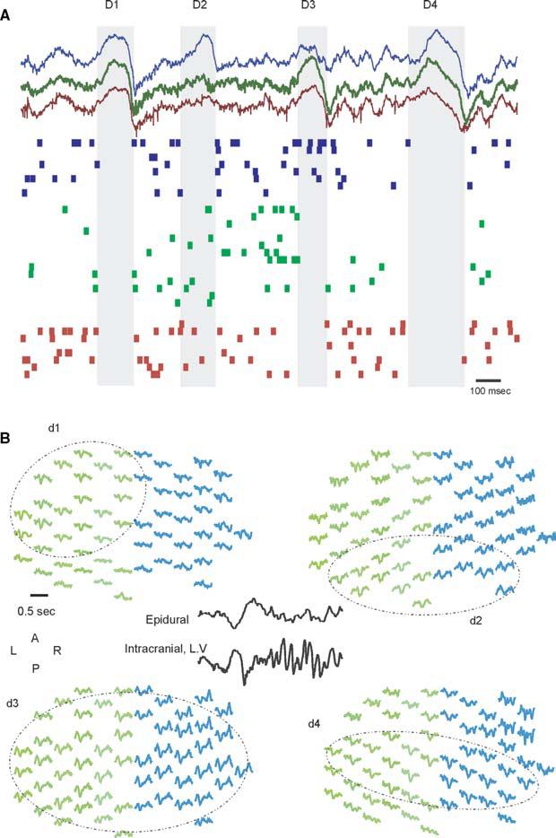
(A) Example of simultaneously recorded LFP and unit activity at three intracortical locations (∼1 mm spaced). Note that DOWN states (shaded area) can be synchronous and global (D1, D4) and local (D2, D3). (B) Example of bilateral, 64-channel, epidural EEG recordings in the rat during SWS. Negativity in the EEG is associated with deep delta wave (inset). Typically, delta waves are globally synchronized (d3), but can be localized to one side (d1), posterior (d2) or central (d4) regions.