
Fig. 1.
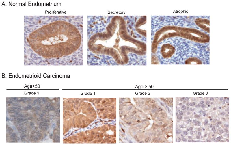
PKCδ expression is reduced in malignant epithelium relative to normal tissue: Representative images of (A) normal proliferative, secretory or atrophic endometrium and (B) endometrioid carcinoma of increasing grade. Samples were processed and stained for PKCδ, as described in Materials and Methods.