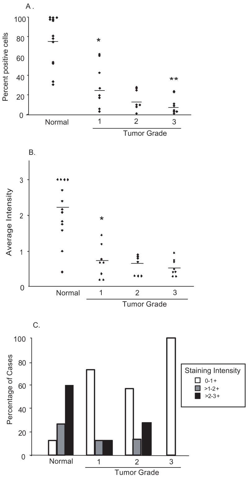
Fig. 4.
Nuclear PKCδ expression is markedly reduced or lost in endometrial carcinomas: Tumor samples were stained and analyzed for (A) the fraction of cells per case exhibiting nuclear PKCδ staining and (B) average staining intensity in the nucleus. The mean percentage stained cells (A) and intensity (B), for each sample population of normal or increasing tumor grade, is denoted by the horizontal line. (C) The fraction of cases falling into each tertile of nuclear staining intensity, according to tumor grade. * p <0.0001. ** p <0.05.

