
Fig. 6.
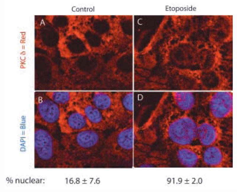
Translocation of PKCδ in response to etoposide-induced apoptosis: HEC-1-A cells were treated with 50μM etoposide or diluent (control), for 6h as indicated. Cells were fixed, permeabilized, probed with antibody to PKCδ and stained by immunofluorescent secondary antibody, as described in Materials and Methods. (A) Control cells. (B) Etoposide treated cells. (C & D) Nuclei counter stained with DAPI. Representative confocal images from two independent experiments are shown. Percent nuclear PKCδ was estimated by counting of five random fields of control or etoposide treated cells, by three independent observers. Results are mean ± s.e.m.