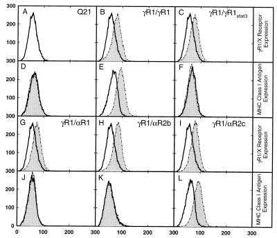
Figure 4.
Expression of receptors on the cell surface and induction of HLA-B7 surface antigen in hamster Q21 cells by IFN-γ. The expression of γR1/γR1, γR1/γR1Stat3, γR1/αR1, γR1/αR2b, and γR1/αR2c (B, C, G, H, and I) or induction of HLA-B7 antigen by IFN-γ (D, E, F, J, K, and L) were analyzed by flow cytometry. (A, B, C, G, H, and I) Cells were harvested and incubated with anti-IFN-γR1 monoclonal antibody (thin lines, shaded areas), and the parental Q21 cells were used as a control (thick lines, open areas). (D, E, F, J, K, and L) Cells were treated with IFN-γ (thin lines, shaded areas) or left untreated (thick lines, open areas). Cells were the parental Q21 cells (A and D) and clonal populations of hamster cells stably transfected with the following: γR1/γR1 (B and E), γR1/γR1Stat3 (C and F), γR1/αR1 (G and J), γR1/αR2b (H and K), and γR1/αR2c (I and L).

