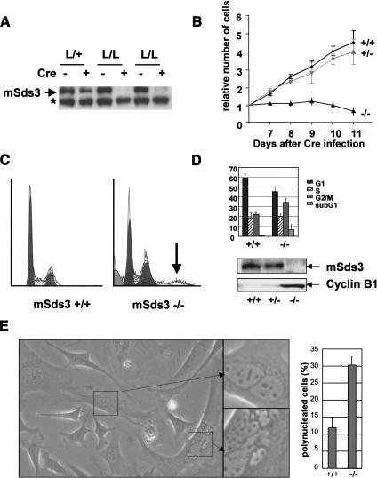
Figure 2.
mSds3 deficiency results in cell cycle defects. (A) Western blot analysis with an anti-mSds3 antibody of MEFs, 8 d after Cre recombinase infection. The asterisk marks a nonspecific band. (B) Growth curve of MEFs wild-type, heterozygous or null for mSds3 after Cre recombinase infection. For each genotype, six independent cell lines were tested for this assay in duplicate. Empty vector-infected mSds3L/L MEFs grew similar to wild-type MEFs (data not shown). (C) Flow cytometry analysis of mSds3 MEFs 8 d after Cre recombinase infection. Shown is a representative result obtained for mSds3+/+ and mSds3-/- cells. The arrow points to polyploid cells in mSds3-/- cell population. (D, top) Cell cycle repartition average on two independent cell lines for each genotype in duplicate 8 d after Cre recombinase infection. (Bottom) Western Blots performed on the indicated cell lines with an anti-mSds3 antibody or an anti-cyclin-B1 antibody 8 d after Cre recombinase infection. (E, left) Bright field picture of representative mSds3-/- MEFs culture 8 d after Cre recombinase infection. (Right) Quantification of polynucleated cells in three independent primary MEFs of each genotype (at least 200 cells were counted for each cell line).

