Figure 6.
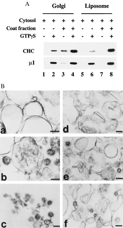
Electron-microscopic analysis of clathrin-coat assembly on Golgi membranes and liposomes. (A) Golgi membranes and soybean 20% PC liposomes were incubated with 5 mg/ml bovine adrenal cytosol supplemented with 15 μg/ml soluble CCV coat fraction, 4 μM mARF1, and 100 μM GTPγS as indicated. The recruitment of clathrin (CHC) and AP-1 (μ1) was determined by immunoblotting. (B) Membrane pellets recovered from Golgi membranes and liposomes that had been incubated as above were fixed and processed for electron microscopy as described in Materials and Methods. Golgi membranes incubated with cytosol and coat fraction in the absence (a) and presence (b) of GTPγS. Liposomes incubated with cytosol and coat fraction in the absence (d) or presence (e and f) of GTPγS. (c) Purified CCVs from rat liver. Clathrin-coat assembly occurred only in the presence of GTPγS (b, e, and f). (Bar = 100 nm.)

