
Figure 6. Expression of selected IFNγ-responsive genes is extended in neurons as compared to MEF after an IFNγ pulse.
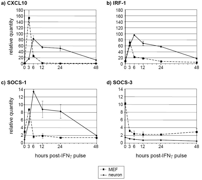
Neurons and MEF were treated as described in Figure 5a above. Total RNA was purified from the lysates and analyzed using RT-qPCR for the presence of CXCL10 (a), IRF-1 (b), SOCS-1 (c), and SOCS-3 (d) transcripts. The timepoints correspond to the length of time in h after the IFNγ was washed out.